
Introduction
A German Shepherd spends hours scrambling over jagged rocks, loose shale, and fallen timber during a wilderness search. The next morning, the dog is reluctant to put weight on one paw. The handler notices subtle swelling between the toes and a sharp reaction when the digital flexor tendons are palpated. This is digital tendonitis—a common yet often overlooked injury in working search and rescue dogs. Left untreated, the condition progresses to chronic lameness, scarred tendon sheaths, and an early end to a hero‘s career. Extracorporeal shock wave therapy (ESWT) has emerged as a powerful treatment for precisely this type of injury, offering a non-invasive way to reduce inflammation, stimulate tendon healing, and return working German Shepherds to the field faster than traditional rest‑only protocols. This article explains how rocky terrain causes digital flexor tendonitis, how ESWT works at the cellular level inside damaged canine tendons, and how handlers can incorporate this therapy into their dog’s rehabilitation plan.
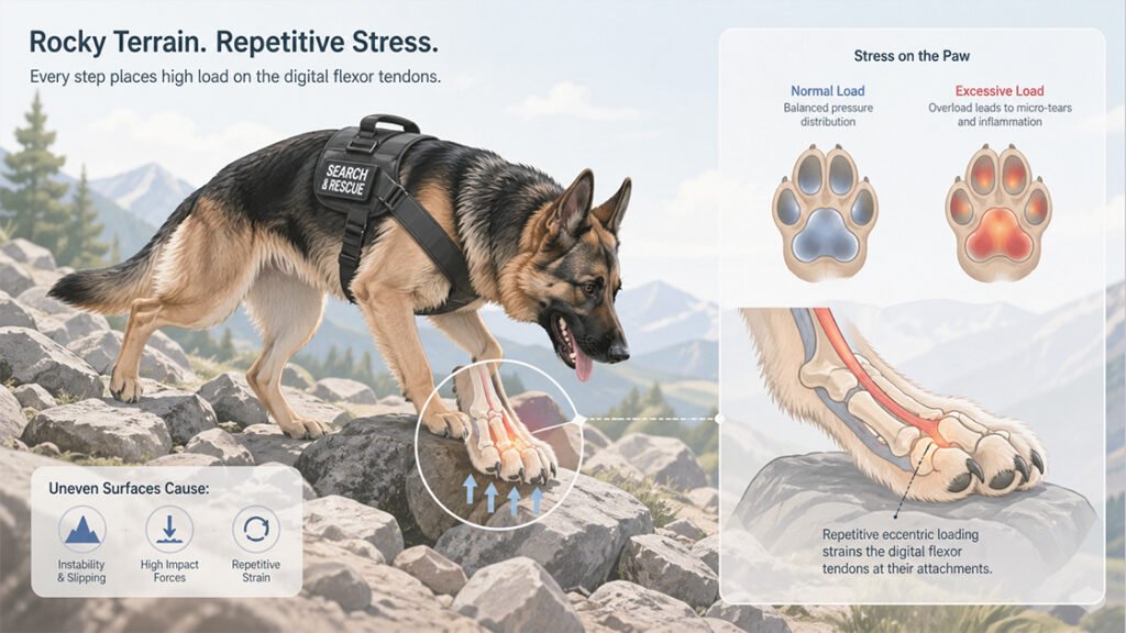
1. Why Rocky Terrain Destroys Working Dog Paws
Search and rescue German Shepherds are bred for endurance, intelligence, and drive. They do not quit even when injured. Unfortunately, the very terrain that makes them invaluable for wilderness recovery also inflicts cumulative trauma on their paws and digits.
1.1 The Digital Flexor Mechanism in Dogs
Each canine digit contains two flexor tendons—the superficial and deep digital flexors—that run from the elbow and carpus down to the toe bones. These tendons slide through synovial sheaths, allowing smooth gliding as the dog grips uneven surfaces. During a rocky terrain search, the dog constantly contracts these tendons to stabilize the paw against loose and shifting substrates. The repetitive eccentric loading strains the tendon fibers at their attachment points to the phalanges. Without adequate rest between searches, micro‑tears accumulate faster than the body can repair, leading to a classic overuse tendinopathy with inflammation, swelling, and progressive pain on weight bearing.
1.2 Why Working German Shepherds Are at High Risk
German Shepherds have a naturally high drive that overrides pain signals. A handler may not notice early signs of digital tendonitis until the injury is well advanced. The breed‘s angulated hindquarters also distribute force differently than other working breeds, concentrating more stress through the rear paw digits during steep descents. In addition, search and rescue missions often occur in unpredictable environments—post‑disaster debris, mountainous scree fields, or snow‑covered rubble. These surfaces cause the paw to slide and the tendons to snap suddenly into a stretched position, creating acute strains on top of chronic wear. Many handlers underestimate how quickly seemingly minor paw soreness can become a career‑ending condition.
1.3 Subtle Signs Handlers Often Miss
Digital tendonitis does not always present as a dramatic limp. More often, handlers notice subtle changes: the dog hesitates before descending a slope, shakes the paw after walking a short distance, or licks between the toes when resting. On examination, gentle pressure along the digital flexor tendons elicits a flinch. The toes may feel warm to the touch, and mild swelling appears between the metacarpal pads in the front foot or metatarsal pads in the hind. Without intervention, the condition progresses to palpable thickening of the tendon sheath—a sign of chronic scarring. By the time the dog is visibly lame, significant tissue damage has already occurred.
2. How ESWT Reaches Deep into the Canine Paw
Extracorporeal shock wave therapy delivers high‑energy acoustic waves that penetrate deep into soft tissue without incisions or needles. For a working dog‘s digital tendonitis, ESWT targets the source of pain and poor healing directly.
2.1 From Kidney Stones to Tendon Healing
ESWT was originally developed in human medicine to break up kidney stones through lithotripsy. Veterinarians later discovered that the same acoustic waves, when delivered at lower energy, stimulate healing in poorly vascularized tissues like tendons. The shockwave generators convert low‑frequency, high‑velocity acoustic waves into mechanical energy that transmits through tissues according to their acoustic impedance.-1 When the wave reaches a tissue interface—such as where the digital flexor tendon attaches to bone at the enthesis—it releases its energy, creating micro‑cavitation bubbles that expand and collapse. This implosion generates shear forces that break down disorganized scar tissue and calcific deposits.-1
2.2 The Cellular Cascade of Healing
Once the shockwave energy reaches the damaged tendon, it triggers a cascade of biological responses. The acoustic disruption causes controlled micro‑trauma that stimulates cells to release anti‑inflammatory cytokines and growth factors into the treated tissues.-11 Specifically, ESWT upregulates vascular endothelial growth factor (VEGF) and transforming growth factor‑beta (TGF‑β), which promote angiogenesis—the growth of new capillaries into the hypovascular tendon core.-2 Improved blood flow delivers oxygen and nutrients to an area that normally heals very slowly. At the same time, the shockwave reduces pro‑inflammatory mediators like substance P and prostaglandin E2, decreasing pain and swelling at the injury site.-1
2.3 Breaking the Pain Cycle Without Drugs
Digital tendonitis in working dogs is notoriously painful because the inflamed tendon sheath entraps the gliding fibers with every step. Oral non‑steroidal anti‑inflammatory drugs (NSAIDs) reduce systemic inflammation but do not alter the local mechanical environment of the scarred sheath. ESWT provides a different approach. The acoustic waves temporarily overload the small nerve fibers that transmit pain signals, a phenomenon called hyperstimulation analgesia. Many dogs show immediate improvement in weight‑bearing after a single session, allowing them to begin appropriate rehabilitation exercises while the deeper tendon healing progresses.-11 Because ESWT is applied directly to the affected area, it avoids the gastrointestinal and renal side effects associated with long‑term NSAID use in working dogs.
2.4 Focused vs. Radial: Which One for Paws
Two types of shockwave exist. Radial pressure waves spread outward from the applicator, covering a wider but shallower area. Focused ESWT concentrates energy at a precise depth, adjustable from 5 mm to 40 mm or more.-2 For digital tendons—which sit just millimeters beneath the paw pad and between the metacarpal bones—focused ESWT allows the veterinarian to deposit therapeutic energy exactly at the enthesis without overheating the surface skin. Focused systems also operate with minimal noise, which matters when treating a sound‑sensitive German Shepherd.-2 Most working dogs tolerate focused ESWT without sedation, giving handlers real‑time feedback on pain localization.
3. Practical ESWT Protocol for Search and Rescue Dogs
A rushed or improperly applied shockwave protocol can waste the handler‘s time and the dog’s comfort. Success depends on correct timing, accurate targeting, and appropriate aftercare.
3.1 When to Start After the Injury
The ideal window for ESWT is the subacute phase, roughly 48 to 72 hours after the last search, once the initial inflammatory surge has peaked. Treating an acutely swollen, fresh injury may cause excessive pain and edema. However, delaying too long—beyond two weeks of persistent lameness—allows disorganized scar tissue to mature, making it harder to remodel. For a German Shepherd that shows ongoing digital tenderness after 72 hours of rest, schedule the first shockwave session immediately. Do not wait for the dog to “get better on its own.” Tendons seldom heal without active intervention.
3.2 Targeting the Digital Flexor Enthesis
The veterinarian first palpates the affected paw to localize the point of maximal tenderness—usually just proximal to the synovial sheath of the digit. The area is clipped and ultrasound gel is applied.-3 Using a focused ESWT handpiece, the veterinarian delivers 800 to 1500 pulses at an energy flux density of 0.10 to 0.18 mJ/mm². The frequency is set between 4 and 8 Hz. The veterinarian moves the probe slowly over the digital flexor tendon, from the accessory carpal pad distally to the insertion on the distal phalanx. The session lasts approximately 8 to 12 minutes. Most dogs tolerate awake treatment without sedation; the sensation is a deep tapping rather than sharp pain.
3.3 Number of Sessions and Spacing
A typical protocol for digital flexor tendonitis involves three sessions spaced 10 to 14 days apart. Some cases of chronic enthesiopathy may require a fourth session after three to four weeks. The goal is to stimulate neovascularization and collagen remodeling gradually rather than overwhelming the tendon with excessive energy. Between sessions, the dog should remain confined to short, leashed walks for elimination only. No running, jumping, or free play on rough surfaces is allowed during the treatment series. Handlers often report noticeable reduction in paw‑shaking and improved willingness to bear weight after the second session. Full resolution of lameness typically occurs two to three weeks after the final pulse.
3.4 What Not to Do After Each Session
For 48 hours after ESWT, avoid any anti‑inflammatory medications including NSAIDs and corticosteroids. These drugs blunt the very inflammatory signal that shockwave relies on to trigger healing.-11 Ice packs and cold therapy should also be avoided for the same reason. Do not allow the dog to resume normal search training until cleared by the rehabilitation veterinarian. The potent analgesic effect of shockwave can temporarily mask pain for up to 48 hours, tempting the handler to push the dog too soon.-11 Working dogs that return to rocky terrain within the first week after treatment risk re‑injuring the tendon before remodeling is complete.
4. Long‑Term Paw Health for Working German Shepherds
One episode of digital tendonitis often indicates that the dog‘s paws have taken multiple smaller insults over many missions. ESWT not only treats the current injury but also reduces the likelihood of recurrence when integrated into a maintenance plan.
4.1 Maintenance Sessions Between Deployments
After completing the initial three‑session protocol and allowing four weeks of healing, many working German Shepherds benefit from a single‑session maintenance treatment every four to six months. This preventive ESWT targets subclinical tendon thickening before it becomes symptomatic. A 2022 systematic review of ESWT in dogs, horses, and cats confirmed positive effects in conditions affecting ligaments and tendons, with maintenance protocols emerging as a standard practice for elite working animals.- Handlers who incorporate biannual paw exams with focused shockwave report fewer missed deployments due to chronic lameness.
4.2 Boots and Terrain Management
No therapy replaces smart prevention. After ESWT, fit the dog with high‑quality canine boots that provide shock absorption and abrasion resistance for rocky terrain. Rotate between two sets of boots to ensure they dry completely between uses, preventing moisture‑associated dermatitis that mimics digital pain. Gradually condition the paws to rough surfaces, starting with short sessions on packed gravel before progressing to loose shale. Teach the dog to avoid jumping from heights that jar the digital joints. These environmental modifications extend the benefits of shockwave therapy substantially.
4.3 When ESWT Is Not Enough
Severe digital tendonitis with ultrasound evidence of complete tendon tearing, deep calcification within the sheath, or septic tenosynovitis may not respond to ESWT alone. Signs that the injury exceeds conservative treatment include a draining tract from the paw, fever, marked joint effusion of the distal interphalangeal joint, or failure to improve after two consecutive shockwave sessions. In these cases, surgical exploration with tenoscopy and sheath debridement becomes necessary. However, for the vast majority of grade 1 and grade 2 digital flexor tendinopathies—overuse injuries without visible tearing—ESWT resolves lameness completely and returns the German Shepherd to the search field.
Frequently Asked Questions (FAQ)
Q1: Will ESWT hurt my dog during the session?
Most dogs feel a tapping sensation but remain comfortable without sedation. Focused shockwave devices operate quietly and deliver energy precisely, minimizing discomfort.
Q2: How soon can my German Shepherd go back to searching after ESWT?
Light leashed walks for elimination are allowed immediately. Full‑duty search work typically resumes four to six weeks after completing the three‑session protocol.
Q3: Do I need to clip the paw fur before ESWT?
Yes. The veterinarian will clip the hair over the digital flexor tendon to prevent air pockets that reduce shockwave energy delivery.
Q4: Can I combine ESWT with other therapies?
Yes. ESWT works well alongside underwater treadmill training, therapeutic ultrasound, and targeted stretches once the acute pain resolves.
Conclusion
A search and rescue German Shepherd’s paw takes thousands of punishing steps across rocky terrain that no boot can fully protect. Digital flexor tendonitis is the quiet toll of those missions—a slow buildup of micro‑scarring that eventually robs the dog of a pain‑free gait. Extracorporeal shock wave therapy offers a targeted, drug‑free solution. By delivering focused acoustic energy to the tendon enthesis, ESWT breaks down scar tissue, stimulates new blood vessel growth, and reduces inflammation without the side effects of long‑term medication. A typical course of three sessions returns most working dogs to full field duty within weeks, not months. Shockwave does not replace thoughtful handling and proper paw conditioning, but it ensures that when the call comes, your German Shepherd’s paws are ready to answer.
References
Shockwave Machines. Indications for Shockwave Therapy.
https://www.shockwavemachines.com/indications
PulseVet. Shock Wave & PEMF Therapy for Dogs: Evidence‑Based Rehabilitation Protocols.
https://pulsevet.zomedica.com/shock-wave-pemf-therapy-dogs-veterinary-rehabilitation/
Redding Horne C, Schnabel LV. Extracorporeal shock wave therapy in equine and canine practice. J Am Vet Med Assoc. 2025.
https://avmajournals.avma.org/view/journals/javma/aop/javma.25.12.0827/javma.25.12.0827.xml
dvm360. Why shock wave therapy can’t be imitated in patient care.
https://www.dvm360.com/view/why-shock-wave-therapy-can-t-be-imitated-in-patient-care
Leeman JJ, Shaw KK, Mison MB, et al. Extracorporeal shockwave therapy and therapeutic exercise for supraspinatus and biceps tendinopathies in 29 dogs. Vet Rec. 2016;179(15):385.
https://pubmed.ncbi.nlm.nih.gov/27444781/
Tsai FC, Alvarez LX. Outcome of eight working dogs with fibrotic myopathy following extracorporeal shockwave and rehabilitation therapy: a case series. Front Vet Sci. 2024.
https://www.frontiersin.org/journals/veterinary-science/articles/10.3389/fvets.2023.1258319/full